

43
likes
9
comments
daily view 0
monthly view 0
Live Analytics
Comments 0
Inferior Vena Cava: Where Surgeons Go Wrong | Step 1 Analytics Table
Income Estimates for Inferior Vena Cava: Where Surgeons Go Wrong | Step 1
Based on this YouTube video's total view count of 2.32K views and industry-standard rates, the estimated total earning is $2 - $5 through ad revenue. Historical data is not yet available to calculate daily, weekly, or monthly averages.
About Inferior Vena Cava: Where Surgeons Go Wrong | Step 1
Explore Inferior Vena Cava: Where Surgeons Go Wrong | Step 1 with 2,327 views, 43 likes, and 9 comments. Experience the impact of this video content that has captured audience attention.
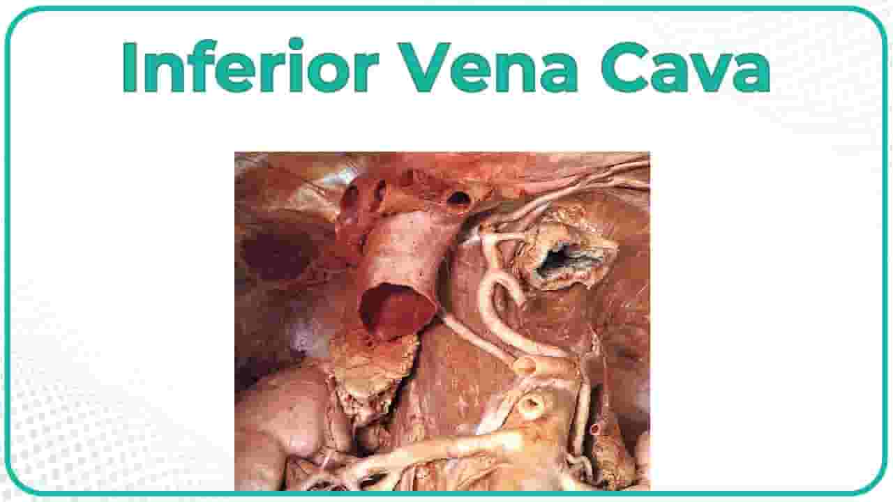
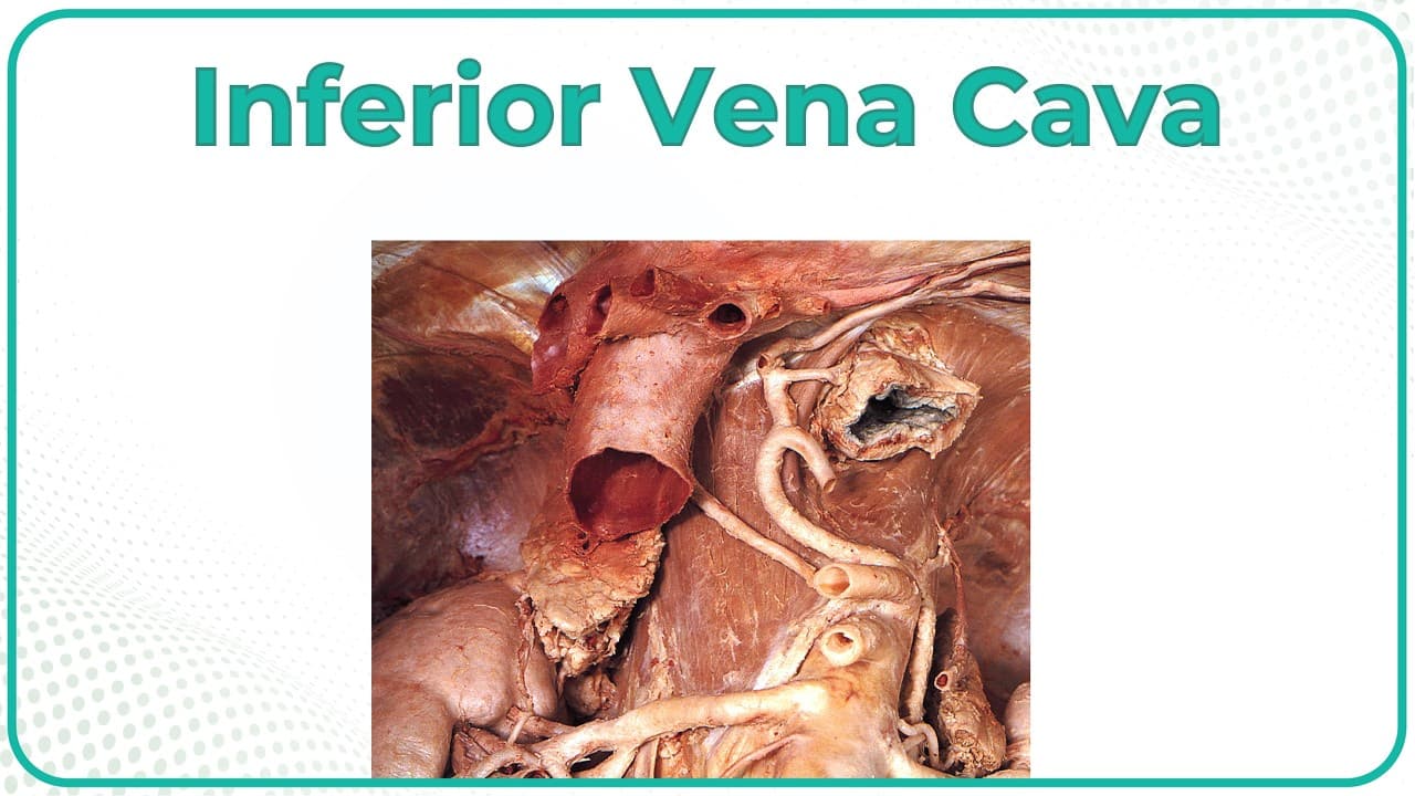
📌𝗝𝗼𝗶𝗻 𝗢𝘂𝗿 𝗧𝗲𝗹𝗲𝗴𝗿𝗮𝗺 𝗖𝗵𝗮𝗻𝗻𝗲𝗹 𝗛𝗲𝗿𝗲:- https://t.me/bhanuprakashdr 📌 𝐅𝐨𝐥𝐥𝐨𝐰 𝐨𝐧 𝐈𝐧𝐬𝐭𝐚𝐠𝐫𝐚𝐦:- https://www.instagram.com/drgbhanuprakash 📌𝗦𝘂𝗯𝘀𝗰𝗿𝗶𝗯𝗲 𝗧𝗼 𝗠𝘆 𝗠𝗮𝗶𝗹𝗶𝗻𝗴 𝗟𝗶𝘀𝘁:- https://linktr.ee/DrGBhanuprakash Inferior Vena Cava (IVC) | Cadaveric Anatomy, Relations & Clinical Correlations | USMLE Step 1 In this cadaveric anatomy demonstration, Dr. G Bhanu Prakash explores the Inferior Vena Cava (IVC), the largest vein in the human body and a critical vessel for systemic venous return. Formed at the level of L5 vertebra by the union of the common iliac veins, the IVC ascends through the abdomen to drain into the right atrium of the heart. Its course and relations make it one of the most important retroperitoneal structures to identify during cadaveric dissection, especially for USMLE Step 1. On cadaveric specimens, the IVC lies to the right of the abdominal aorta, running superiorly through the abdomen and piercing the diaphragm at the level of T8 via the caval opening. Along its path, it receives multiple tributaries, including the hepatic veins, renal veins, right suprarenal vein, right gonadal vein, and lumbar veins. The asymmetry of venous drainage is best appreciated in dissection: the right gonadal and suprarenal veins drain directly into the IVC, while their left counterparts drain into the left renal vein before joining the IVC. The IVC’s close anatomical relations are important in surgery and radiology. It runs posterior to the liver, adjacent to the pancreas, duodenum, and right kidney, making it vulnerable in retroperitoneal pathologies. On cadaveric view, its broad caliber and thin walls make it distinguishable from the aorta, which has thicker muscular walls. Clinically, the IVC is significant in conditions such as deep vein thrombosis (DVT) with risk of pulmonary embolism, IVC obstruction, and tumor invasion (e.g., renal cell carcinoma spreading into the renal vein and IVC). Surgeons may also place IVC filters to prevent emboli from reaching the lungs. By integrating cadaveric identification, tributaries, anatomical relations, and clinical relevance, this lecture provides medical students, residents, and clinicians with exam-ready insights for USMLE Step 1 and practical surgical anatomy orientation. #InferiorVenaCava #CadavericAnatomy #IVCAnatomy #USMLEStep1 #GrossAnatomy #ClinicalAnatomy #Retroperitoneum #USMLEPreparation #MedicalStudentsUSA #DoctorEducation #Step1Prep #CadaverDissection #AnatomyLecture
About YouTube Real-Time View Count
With SocialCounts.org’s view counter, track your YouTube video’s live view count and YouTube likes count in real time with fast, reliable updates.
Watch every YouTube video live view count rise with our real-time YouTube views tracker—built for accuracy and minimal delay.
Follow YouTube real time views as they happen, using our dedicated view counter for YouTube videos.
Get up-to-date live view count on YouTube and see real-time growth with SocialCounts.org’s smart tracking tools.
Embed Widget
Parameters:
fullscreen=true- Fullscreen countergraph=true- Live graph chartcounter=0/1/2- Select counter (0=likes, 1=views, 2=comments)
URL
Click to copy the embed URL to your clipboard